No more boundaries for radiology images
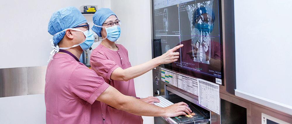
Radiology images are essential to accurate clinical diagnosis and effective treatment. Since April 2015, Hospital Authority (HA) has launched the Filmless Operating Theatres (Filmless OT) Project and introduced the technology in 23 hospitals, with over 230 operating theatres across all seven clusters. Not only does the project allow quick and easy access to digital radiology images for knowing better of patients’ conditions, but it also re-engineers the clinical practices, especially pre-operative planning, to enhance service quality and patient safety.
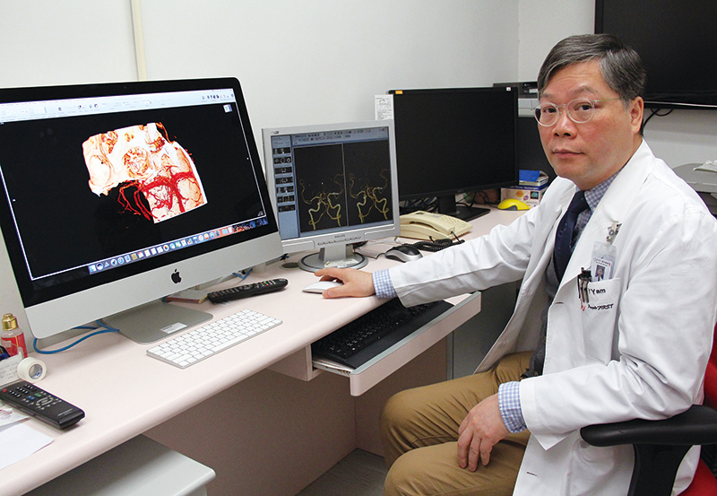
“Digitalised imaging transcends geographical constraints and simplifies the process of image acquisition for better inter-departmental or hospital communication,” says Dr Yam Kwong-yui, Chairman of the Filmless OT Project Advisory Group and Chief of Services of Department of Neurosurgery in Tuen Mun Hospital. He explains that patients might be transferred to another hospital after radiology examination. Before the implementation of Filmless OT Project, patients had to bring along with the physical imaging films for surgery and follow-up, which was way more time-consuming and subjected to a higher risk of image loss.
Nowadays, clinicians from different specialties can access the radiology images soon after patients’ examination as the digitalised images are all uploaded to the corporate-wide central storage. For those cases that need inter-hospital transfer, clinicians can even retrieve the images for preliminary review during the course of patient transportation.
With the installation and use of image processing system inside and outside operating theatres, clinicians can plan the operation in advance. For example, multi-planar reconstruction and 3D volume rendering are two commonly used functions of the system which turns 2D radiology images into a 3D view. These facilitate clinicians to ’visualise’ the inside of patient’s body, or even have a detailed look at the expansion, contraction or other multi-perspective inspection of the involved operative site. Diameter or volume measurements of an internal organ are other features that support pre-operative planning as well.
Taking cerebrovascular aneurysm as an example, Dr Yam explains how the image processing system facilitates clinicians to preview and simulate the operation. It shows clinicians the operative site from multiple angles, and provides a view to seeing the exact size and shape of the aneurysm and all the surrounding branches. As such, surgeons will know and decide which approach (e.g. coiling or clipping) is a better option for the patient. On the day of operation, processed images will be sent and displayed on the viewing equipment inside operating theatres as a reference. In short, clinicians can make a better operative plan with all these fine cut radiology images.
● New technology to enhance patient safety
COVER STORY
● No more boundaries for radiology images
● Development of filmless technology
● Streamlined workflow model for four million sets of radiology images
● Triumph over 9-year uphill struggles
FEATURE
● Restoring the distinct past of KWH
● ‘Like for like’ approach in restoration
PEOPLE
● Young physiotherapist builds a playground in developing country
● Most-loved facilities in the playground
HELEN HA
● Small electrical appliances now available at the online shop
WHAT'S NEW
● New leaders adopt a down-to-earth leadership style
● Mothers’ great companion on the journey of breastfeeding
● Measures to facilitate breastfeeding in workplace
● Make the most out of big data in service planning
● Big data analytics platform to be launched by year-end – no copy and take away of data
STAFF CORNER
● TPH psychiatric rehabilitation centre develops multi-disciplinary training for patients’ recovery
● Fall prevention programme helps elders stay safe and healthy
● 我們都在這裡!(Chinese version only)


