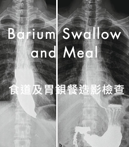
Barium Swallow and Meal 食道及胃鋇餐造影檢查
Indication 檢查目的:
Suspected pathology in the oesophagus or stomach. 檢查病人食道及胃部的病理。
Procedure 方法:
The examination involves the patient taking a suitable amount of oral contrast medium (containing barium compounds). A series of X-rays are then taken. The examination is performed by a radiologist. 病人在放射科醫生指導下進飲適量鋇劑,同時醫生利用X光透視,詳細觀察病人食道及胃部的活動和病變,並同時進行X光攝影。
Possible Complications Of Use Of Contrast Medium 副作用:
- 1. Abdominal discomfort due to distension of the stomach. 鋇劑或會使病人胃部不適。
- 2. Aspiration of contrast medium into the lung. 服食之鋇劑可能吸進肺部而引致氣管不適或其它相關的併發症。
- 3. Leakage of contrast medium due to any unexpected perforation. 如果病人的食道或胃部有穿孔,鋇劑可能流入腹膜而引致相應的併發症。
- 4. Constipation after taking the barium contrast. 鋇劑會導致便秘。
Patient Preparation 檢查前準備:
- 1. Adult patient should fast for 8 hours before the examination. 成年人在檢查前八小時不可飲食。
- 2. Smoking should be discontinued or restricted. 禁止吸煙。
- 3. For paediatric patient under the age of 3 years, fast 3 hours before the examination. 三歲以下小童,在檢查前三小時不可飲食。
- 4. For children between 3 to 12 years of age, fast for 8 hours before examination. Light supper only in the evening before the examination. 三至十二歲小童,在檢查前八小時不可飲食。
- 5. Patient should follow the instruction of the staff during the examination as various positions may be adopted to facilitate the flow of contrast. 病人應與醫護人員充份合作。在檢查過程中,病人身體須作不同角度轉動,以便所服鋇劑能清晰地顯現出整個上消化道的實況。
- 6. Paediatric patients should be accompanied by parents. 兒科病人應由家長陪同前來檢查。
- 7. For diabetic patient on drugs - consult clinician concerned for the adjustment of insulin dosage if necessary. 糖尿病人請遵照醫生指示調節糖尿藥份。
In case of doubt please ask our radiological staff or the referring clinician. Please inform our staff before the examination if you think you are or may be pregnant. 病人如懷疑其本人可能或已經懷孕,在檢查前切記通知本部人員。如有任何疑問,請向主診醫生或放射科醫生查詢。
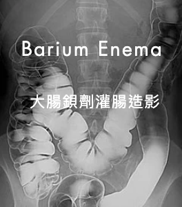
Barium Enema 大腸鋇劑灌腸造影
Indication 檢查目的:
Suspected pathology in the colon. 檢查病人大腸病理。
Procedure 方法:
The examination requires bowel preparations such as cleansing enema before the study. During the examination, contrast medium (containing barium compounds) is introduced into the large bowel via a rectal catheter. A series of X-rays are then taken. The examination is performed by a radiologist. This is an unpleasant examination. Some patient may experience abdominal pain during the introduction of air and contrast agent. Inform the radiologist as soon as possible if you feel any pain. 病人在檢查前須先在病房內接受灌腸清洗程序。檢查時放射科醫生會利用X光透視觀察,將鋇劑和空氣通過導管注入直腸及大腸內,並同時進行X光攝影。當檢查進行時,病人如感覺腹部疼痛或任何不適,應立即通知醫生。
Possible Complications Of Use Of Contrast Medium 副作用:
- 1. Abdominal discomfort due to distension of the large bowel by gas and contrast media. 大腸內注入空氣及鋇劑,會引起腹部脹痛不適。
- 2. Perforation of bowel. 病人腸壁如因近期手術而有輕微破損,鋇劑造影檢查可能導致大腸穿孔而引發 相應的併發症。
- 3. Venous intravasation of contrast. 鋇劑可能滲入靜脈血管內。
- (Complications 2 & 3 may occur only in patients with critical bowel lesions and the incidence is very low) (上述二及三兩項副作用,只是在個別的病例才可能發生,且發生率極微。)
Patient Preparation 檢查前準備:
- 1. Patient should follow the instructions for bowel preparation as good quality films largely depend on adequate bowel preparation. 為確保高質素的X光檢查效果,病人必須遵從醫護人員的指導如下
- a) Maintain a low residual diet (i.e. one restricted to bread, potatoes, rice and other starchy foods, but no vegetables or fruits) for at least 48 hours prior to examination. Fluid diet (e.g. honey, pure soup, tea, water, pure fruit juice, pure jelly, etc.) 24 hours before examination. 檢查前二天低渣滓飲食(如麵包、薯仔、飯等澱粉質食物,但不能吃蔬菜、生果)。
- b) A suitable aperient prescribed by doctor in-charge shall be given on evening before examination. 檢查前一天流質飲食 (如蜜糖水,清湯,清茶,開水,清果汁, 啫喱等)。
- c) Fluid diet on the day of examination and cleansing enema should be performed. 檢查前一晚,依醫生指示服用瀉藥。檢查日流質飲食及要接受灌腸清洗程序。
- 2. Patient should follow the instructions of the staff during the examination as various positions may be adopted to facilitate the flow of contrast.病人在檢查進行中,應與本部門醫生及放射技師合作,病人身體須遵照工作人 員的指引而作不同角度轉動,以配合X光儀器拍攝大腸各部份。
- 3. For diabetic patient on drug - consult clinician concerned for the adjustment of insulin dosage if necessary.糖尿病人請遵照醫生指示調節糖尿藥份。
In case of doubt please ask our radiological staff or the referring clinician. Please inform our staff before the examination if you think you are or may be pregnant.注意:病人如懷疑其本人可能或已經懷孕,在檢查前切記通知本部人員。如有任何疑問,請向主診醫生或放射科醫生查詢。
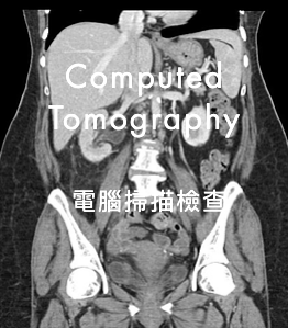
COMPUTED TOMOGRAPHY (CT) 電腦掃描檢查
Indication 檢查目的:
To provide diagnostic information for suspected pathology in specific organs or areas of the body. 診斷病人體內未能確定的病因,對指定的部位或器官作特別的檢查。
Procedure 方法:
During the examination, the patient will be lying on the table of the CT scanner. The table will then carry the patient slowly through the gantry of the scanner and a well-collimated X-Ray beam will pass through the patient. In this way, a number of cross-sectional images of the organs/areas of interest are made. Intravenous or oral contrast medium may be needed to improve the diagnostic quality of the images. You will be advised to keep still during CT scanning and listen carefully to instructions given by our staff. Medication for sedation are sometime administered to paediatric patients in order to obtain images without undue movement. The whole procedure is monitored by a radiologist. 病人需先躺在掃描機檢查床上,在技術人員的操作下被自動送進掃描機的環形通道內,經過X光照射,然後進行一連串經電腦分析的橫切面造影。如有需要,病人在檢查前需服用造影劑,或在檢查中醫生會替病人在靜脈注射造影劑以增加診斷的準確度。兒科病人或需使用鎮靜劑,以保証檢查能順利進行。
Possible Complications Of Use Of Contrast Medium 靜脈注射造影可能引致的副作用:
- 1. Allergic reaction to intravenous contrast medium. 造影劑可能引致過敏反應。
- 2. Pain at injection site during injection of intravenous contrast medium. 注射時,針口位置可能有疼痛的感覺。
- 3. Aspiration of gastric content or oral contrast medium as nausea and/or vomiting may occur. (Fasting is thus needed prior to examination). 若病人出現嘔吐現象,可能將胃內之造影劑吸入肺部。(故病人需要在檢查前禁食)
Patient Preparation 注意事項:
- 1. Your referring doctor will ask you to sign a consent form if intravenous injection of contrast medium is indicated. You should volunteer information to your doctor on your previous history of:
- (i) allergy to food and drugs, and in particular any previous reaction to contrast medium,
- (ii) asthma, urticaria, eczema and allergic rhinitis etc. 如果在檢查時要為病人作靜脈注射造影,主診醫生會要求病人簽一張同意書。病人即應向主診醫生詳細提供是否有過敏反應的病歷。例如哮喘、風疹、濕疹、過敏性鼻炎等,又或會否對某些食物、藥物(特別是X光造影劑)或任何物質有過敏的反應等。
- 2. For examinations with intravenous contrast medium, no food or drink should be taken at least 4 hours prior to examination.若檢查時需作靜脈注射造影,病人在檢查前四小時不得飲食。
- 3. Please be punctual at our CT department.請準時到達本部。
- 4. Some examinations will take a longer time than the others. e.g. patient having abdominal CT scan might need to take oral contrast medium some hours before the scanning, so please allow sufficient time (about half a day) for the examination. For some in-patients, oral contrast would have to be taken in the ward.檢查所需時間視乎個別情況而定。例如大部份接受腹部檢查的病人需要在檢查前數小時開始飲用口服造影劑,所以請病人耐心等候,並預備充份時間(大約半天)以便檢查能順利完成。
- 5. For diabetic patient on drug - consult clinician concerned for the adjustment of insulin dosage if necessary.糖尿病人請遵照醫生指示調節糖尿藥份。
In case of doubt please ask our radiological staff or the referring clinician. Please inform our staff before the examination if you think you are or may be pregnant.注意:病人如懷疑其本人可能或已經懷孕,切記在檢查前通知本部人員。如有任何疑問,請向主診醫生或放射科醫生查詢。
MAGNETIC RESONANCE IMAGING (MRI)磁力共振掃描
Principles And Indications 磁力共振掃描的原理和應用:
Magnetic Resonance Imaging (MRI) makes use of a strong superconducting magnet, radio waves and computer system to form pictures or images of the body. It provides diagnostic information for suspected pathology in specific organs or areas of the body particularly in brain, spine, joints and soft tissue of extremities. A large part of our body is composed of water which contains hydrogen atoms. These atoms are slightly magnetic. By using a combination of magnetic fields and radio waves, we are able to obtain signals from them. These signals are then converted by the computer into detailed images of various structures of the body. 磁力共振掃描利用磁場原理,再透過電腦,將人體內部器官顯示成為圖像。這一種先進的醫學影像技術,能夠幫助診斷病人體內未能確定的病因,特別適用於腦部、脊骨、關節及四肢的軟組織。 人體細胞主要是由水份組成,在強力的磁場和電磁波的相互作用下,我們可取得水分內氫原子反應所得的訊號,再經由電腦的處理,便可獲得身體內部組織的精細圖像。
Procedure 方法:
The entire examination will normally take about one hour, and occasionally it may take longer. You will hear a regular drumming noise during the scan. Although the actual scanning procedure occurs intermittently, you must keep still and relax throughout the examination. Our MRI staff will be in constant touch with you during the examination time, and you can talk to our staff. In the scan room, you will lie awake on a sliding couch inside the magnet. You will not feel anything or any discomfort. Blankets will be provided to keep you warm and comfortable; and you may also have a pulse monitor and / or electrocardiogram sensor attached. You may receive an injection of an MR contrast medium, a colourless liquid, that enables the radiologist to better see the structures being studied and to better diagnose your condition. You can eat or drink as usual after the examination. The result of your scan will be communicated to your referring doctor. 雖然掃描的過程是間歇性地進行,可是您亦應盡量保持鬆弛和避免作任何身體的移動。當掃描進行時,儀器會發出噪音,病人須戴上耳塞。兒童病人可能需用鎮靜劑來確保安靜,以便獲得清晰的影像。在檢查過程中,我們的工作人員會與您保持聯絡,而您亦可以與他們對話。 在掃描室內,您會舒適地躺在特備的床上,並視乎接受檢查的部位,緩緩地被推進掃描器中央。整個磁力共振檢查約需一至二小時,在檢查進行中,您必須保持安靜,並與醫護人員合作。 在掃描過程中,病人或需要接受靜脈造影劑注射作較長時間的檢查,以協助正確診斷。整個檢查過程會由放射診斷科醫生監察進行。在掃描完畢後,您便可以如常進食。我們會將有關的檢查結果轉達您的主診醫生。
Complications And Contra-Indications 副作用及注意事項:
MRI is safe and at present there are no known hazards. MR contrast medium, gadolinium-DTPA, has been used safely in millions of cases. Minor reactions (principally headache or nausea) occur in about 2% of persons. Serious life-threatening reactions are rare, and have only been reported in about one in 400,000 persons. However, NOT everyone can have an MRI scan because of the effects of strong magnetic field. 磁力共振掃描是先進的影像技術,對病人絕少引起不良反應。檢查所用的造影劑甚為安全,但也可能會引起頭痛、嘔吐、噁心等反應,嚴重的副作用極為罕見。由於磁場的效應,請留意以下情況:
- 1. Patient with cardiac pacemaker / certain artificial heart valve cannot have this type of scan.磁力共振檢查會對安裝在病人體內的一些醫療儀器,例如心臟起博器、人工心瓣等造成干擾,故此類病人不宜接受這項檢查。
- 2. Patient with ferromagnetic surgical clips in the head, metal fragments or metal implants in the eye or ear, artificial joint, skull plates, dentures, metal splinters and permanent eyeliners may not be suitable for an MRI examination.磁力共振檢查可能對病人體內安置的金屬器械造成移位、鬆脫等現象,所以醫護人員會根據有關安全守則,在檢查前向病人詳細查詢。
- 3. It is not advisable for a woman within the first three months of pregnancy to have an MRI scan.雖然目前並未有孕婦因磁力共振檢查而使胎兒受影響的個案紀錄,但在首三個月懷孕期內的孕婦,應該盡量避免這項檢查。
Patient Preparation 檢查前的準備:
Because of the necessity of preparation and the variable examination time for different patients, please come on time for the appointment and allow four hours of stay in the MRI department for completion of the examination. 為確保我們有充足的時間替全部病者進行各種步驟和準備,每位病人務必在約定的時間準時到達,同時亦應預計可能需要在掃描中心逗留長達四小時。在掃描前,請留意以下事項:
- 1. You should NOT eat or drink for 4 hours before the examination. For diabetic patient on drugs - consult clinician concerned for the adjustment of insulin dosage if necessary. 檢查前四小時不可飲食。糖尿病人請遵照醫生指示調節糖尿藥份。
- 2. Our MRI staff will help you complete a questionnaire so as to ensure that you can have an MRI scan.醫護人員會幫助病人填寫一份問卷,以確使病人能在安全情況下進行掃描。
- 3. Lockers are provided for temporary storage of your belongings. However, you are advised not to bring along valuable belongings or large objects.雖然我們會提供暫貯物品的貯物柜,病人亦不應攜帶貴重或體積過大的物品。
- 4. You will be asked to change into a hospital gown. You must remove all metallic objects (e.g. keys, jewellery, watches, pins, eyeglasses and hearing aids). Credit cards or MTR tickets must also be left in the locker as the magnetic data will be erased by the magnetic field.身上所有金屬物品必須除下,對磁場有反應的物件,如信用咭、提款咭、儲值車票等都不能帶進檢查室。
- 5. Patients attending for head scans should use minimal make-up, especially eye make-up, as it will show on the images.接受眼部及腦部檢查的病人,不可有眼部化妝;至於面部化妝,亦必須盡量減少。
- 6. Paediatric patients would require accompanying by parents or guardians. 兒童病人須由家長或親人陪同。
In case of doubt please ask our radiological staff or the referring clinician. Please inform our staff before the examination if you think you are or may be pregnant.注意:病人如懷疑其本人可能或已經懷孕,切記在檢查前通知本部人員。如有任何疑問,請向主診醫生或放射科醫生查詢。
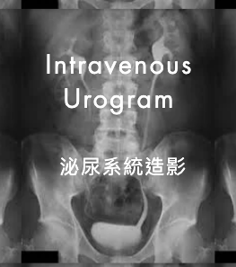
INTRAVENOUS UROGRAPHY 成人泌尿系統造影
Indication 檢查目的:
Suspected urinary tract pathology. 檢查病人泌尿系統病理。
Procedure 方法:
The examination involves the intravenous injection of a contrast medium, after which a series of X-Ray films are taken. Abdominal compression may be applied during the examination. The procedure will be monitored by a radiologist and it usually takes about 1 hour to complete. 將適量的X光造影劑注入病人的靜脈,然後利用X光進行一系列的攝錄。攝影時一般需要利用氣囊緊壓病人下腹,以求取得較好的造影效果。此項檢查大約需要一小時,整個過程由放射科醫生監督進行。
Possible Complications Of Use Of Contrast Medium
- 1.Contrast reactions, such as headache, nausea, vomiting and respiratory distress. 靜脈注射的X光造影劑可能引起副作用(如嘔吐、噁心或呼吸困難)。
- 2.Pain at site of injection. 靜脈注射位置或會引起疼痛。
- 3.Abdominal compression may cause discomfort or hypotension. 下腹部受壓時,可能引起不適或低血壓。
Please inform the radiological staff immediately if any complication occurs. 病人如遇上述情況,請立刻通知在場的放射技師或醫生。
Patient Preparation 檢查前的準備:
- 1. Your referring doctor will ask you to sign a consent form for this investigation since a contrast agent is needed. You should volunteer information to your doctor on your previous history of allergy to food or drugs, history of asthma, urticaria, eczema and allergic rhinitis, and particularly any history of allergy to contrast medium. 主診醫生會預先要求病人簽署同意書。這時病人應向主診醫生詳細說明其本人曾否 對某種食物或藥物產生過敏反應;以及病人曾否患有哮喘、風疹、濕疹、敏感性鼻炎等疾病,最重要是說明病人可曾對X光造影劑有過敏性反應的紀錄。
- 2. Bowel preparation - a suitable aperient prescribed by doctor in-charge shall be given for two successive nights prior to examination to promote bowel action. 檢查前二天,請依照醫生指示,服用瀉藥以便排清大便,有利泌尿系統造影檢查。
- 3. No food for 4 hours before examination, but water may be allowed. 檢查前四小時不可進食,但可以進飲開水。
- 4. If possible, patient should be ambulant for 2 hours prior to examination to reduce bowel gas. 病人盡可能在檢查前二小時起床坐立或走動,減少腸中氣體積聚,使造影有更佳效果。
- 5. Patient with uraemia or myelomatosis should avoid dehydration.病人如呈有尿毒或骨髓瘤病症狀,應多喝開水。
- 6. Patient should empty his/her bladder before the examination. 檢查前病人應排清小便。
- For diabetic patient on drug - consult clinician concerned for the adjustment of insulin dosage if necessary. 糖尿病人請遵照醫生指示調節糖尿藥份。
In case of doubt please ask our radiological staff or the referring clinician. Please inform our staff before the examination if you think you are or may be pregnant.注意:病人如懷疑其本人可能或已經懷孕,切記在檢查前通知本部人員。如有任何疑問,請向主診醫生或放射科醫生查詢。
INTERVENTIONAL RADIOLOGY (IR) 醫療造影服務
Interventional Radiology is a rapidly expanding subspecialty in the field of radiology during today's era of Minimally Invasive Therapy. The procedures are often performed by radiologist through small wounds under ultrasound or X-ray guidance in radiology examination room.
There are various types of procedures with wide range of applications. For additional patient information leaflets on various interventional radiology procedures, please refer to HKSIR web site.
Click for further information on our dedicated IR page.
ANGIOGRAPHY 血管造影檢查
Indication 檢查目的:
To provide diagnostic information for suspected vascular pathology in specific organs or areas of the body. 對指定的部位或器官作特別檢查,協助診斷人體內血管各種病變和病徵。
Procedure 方法:
The examination involves percutaneous introduction of an angiocatheter to a relevant vessel. The angiocatheter is usually introduced via the right femoral artery at the groin under local anaesthesia. A thin catheter is then introduced into the femoral artery through the use of a needle and guide wire. Other sites however may be used. Such as a brachial approach if both femoral pulses are absent. A suitable contrast is then injected and a number of X-Ray images are recorded. Sedation or general anaethesia is usually used for paediatric patients. The examination is performed by radiologist. Make sure you have emptied your bladder before the examination which may take one or sometimes two hours. 檢查時醫生會在病人右腹股溝間進行局部痳醉,切開小部份表皮,插入導管,連接右股上的大動脈,通過透視,將導管移動至與所需檢查的血管接合,然後注入造影劑。整個過程也要用特別X光儀器錄取血管造影。有時可能在前臂彎的位置插入導管。兒科病人可能使用鎮靜劑或全身痳醉。檢查需要一至二小時,病人最好在檢查前排清小便。
After the removal of catheter, the radiologist may need to press the puncture site for about 10 minutes for haemostasis. 當導管被拔出後,醫生將在傷口部位按壓十分鐘以上,幫助止血。
Overall complications are rare. After the procedure, you will still be under observation in the ward. You should inform the attending doctor or nurse as soon as possible in case of any problem. 病人在檢查完畢後,須回病房休息及接受觀察。病人如發覺有任何不適,請盡快通知當值醫生或護士。
Possible Complications
- 1. Contrast reaction.造影劑可能產生副作用。
- 2. Haemorrhage or haematoma at the puncture site.傷口可能因感染而腫痛或流出血水。
- 3. Infection-painful swelling and discharge from the puncture site.可能引致血管栓塞。
- 4. Thrombosis of the vessel.病人表皮因切孔關係,可能有出血現象。
- 5. Embolism.在血管中可能凝成小血塊,導致栓塞。
- 6. Damage to local structures.對傷口附近的組織可能引起破壞。
Patient Preparation 檢查前的準備:
- 1. Your referring doctor will ask you to sign a consent form for this investigation since a contrast agent is needed. You should volunteer information to your doctor on your previous history of allergy to food or drugs, history of asthma, urticaria, eczema and allergic rhinitis, and particularly any history of allergy to contrast medium. 主診醫生會預先要求病人簽署同意書。這時病人應向主診醫生詳細說明其本人曾否 對某種食物或藥物產生過敏反應;以及病人曾否患有哮喘、風疹、濕疹、敏感性鼻炎等疾病,最重要是說明病人可曾對X光造影劑有過敏性反應的紀錄。
- 2. Check any bleeding tendency and correct if possible.檢查血液凝結率是否符合標準,如有問題,須先矯正。
- 3. Fast for 6 hours before the examination.檢查前六小時不可飲食。
- 4. Skin preparation of the puncture site.先將手術部位皮膚上的體毛剃光及清潔,作手術前準備。
- 5. During the examination, you are advised to listen carefully to the instructions given by our staff.檢查進行時,病人應聽從醫護人員指導。
- 6. For diabetic patient on drug - consult clinician concerned for the adjustment of insulin dosage if necessary.糖尿病人請遵照醫生指示調節糖尿藥份。
In case of doubt please ask our radiological staff or the referring clinician. Please inform our staff before the examination if you think you are or may be pregnant.注意:病人如懷疑其本人可能或已經懷孕,切記在檢查前通知本部人員。如有任何疑問,請向主診醫生或放射科醫生查詢。
MAMMOGRAPHY 乳房X光檢查
Indication 檢查目的:
- 1. Screening for breast cancer. 預防性乳癌普查。
- 2. Suspected pathology in the breast. 乳房病徵的檢查。
Patient Preparations:
- 1. Informed consent and allergy History Form are required for Ductogram. 病人如懷疑自己已經懷孕,在檢查前應通知本部門人員。
- 2. Please inform our staff before the examination if you think you may be pregnant. 病人在檢查前須自行將乳房及腋窩部位洗抹乾淨,不可有紗布、除體臭噴劑及爽身粉等物,因上述物品可能影嚮X光片影像的清晰度,妨礙正確診斷。
- 3. Dressing, deodorant (spray, roll-on) agents, talcum powder must not be applied to the breast and axillary region. The films taken with this dedicated machine are very sensitive in picking up skin artifacts which may affect the interpretation of the films.
- 4. Clothing : Blouse, skirt and trousers are preferred. 衣著 : 請勿穿連身裙。
Procedures:
- 1. Mammography is X-Ray imaging of the breast. It involves compression of the breast between a plastic paddle and an X-ray film holder. 透過X光造影方法,錄取乳房內部組織。拍攝時須用膠板把乳房盡量壓平,以取得良好效果。
- 2. Some patients may feel pain or discomfort during compression of their breasts, especially when performed in pre-menstrual period. However, this process is absolutely important because: 病人因乳房受壓可能引致不舒服或疼痛(特別在經期前進行),但此過程非常重要,原因如下 :
- a. Improve image quality, hence increase diagnostic accuracy. 可令X光片影像更加清晰,從而增加診斷的準確性。
- b. Reduce radiation dose. 減少幅射劑量。
- c. Abstaining from coffee or postponing the procedure to post-menstrual period may decrease the discomfort. 檢查前戒飲咖啡,或把檢查改至經期後進行可能會令不適程度降低。
In case of doubt please ask our radiological staff or the referring clinician. Please inform our staff before the examination if you think you are or may be pregnant.注意:病人如懷疑其本人可能或已經懷孕,切記在檢查前通知本部人員。如有任何疑問,請向主診醫生或放射科醫生查詢。
ULTRASOUND 超聲波掃描檢查
What Is An Ultrasound Examination? 甚麼是超聲波掃描?
Ultrasound is high frequency sound wave, too high for human ear to hear. It can pass through the skin and tissues without being felt or heard. Ultrasound examinations use these high frequency sound waves to produce images of tissues in the body. 超聲波是一種高頻音波,它的頻率遠高於人類可以聽見的範圍。它可以穿透人體皮膚和組織。超聲波掃描利用這種音波,經過高解度影像技術,用以檢視人體內部器官。
Why Do I Need Ultrasound Examinations? 為何需要超聲波掃描?
Ultrasound examinations can help doctors detect many diseases. Ultrasonography of the gallbladder may show stones. An image of the breast, thyroid, liver, kidney, or pancreas may show masses. Ultrasound examination of pregnant mother can help doctor to assess the condition of the baby inside the womb. Ultrasonography of major blood vessels can detect problems such as disease or clots. Under normal clinical practice ultrasound examination is painless and harmless to our body. 超聲波掃描可以檢視人體內部的主要器官,例如:肝臟、膽囊、胰臟、腎臟、子官、卵巢和乳房等。與此等器官相關的疾病和症狀,如肝硬化、膽石、胰臟炎、腎石及腫瘤等,都可以準確地診斷出來。孕婦接受超聲波掃描更可檢查胎兒內臟結構及成長。超聲波又可用作檢查主要血管的狀況,顯示血管狹窄、栓塞等。 超聲波掃描在正常情況使用下是一種不含輻射,無傷害性的檢驗,亦不會引起痛楚。
How Do I Prepare For My Exam? 檢查前的準備:
You may be told not to eat 6 hours before ultrasound examination of the liver and gallbladder. For some examinations, you may need to drink several glasses of water beforehand so that your bladder is full during ultrasonography. There may not be any preparation required for other examination regions. You will be given instructions about how to prepare for your examination. For diabetic patient on drug - consult clinician concerned for the adjustment of insulin dosage if necessary.
- 1. 上腹部檢查(如肝、膽等):檢查前六小時須禁止進食。 下腹部檢查(如腎、婦產科等):病人須多喝開水,令膀胱漲滿,以便檢查。 其他部位檢查(如乳房、甲狀腺等):病人無須特別準備。 詳情請參考預約時間通知書上的指示。
- 2. 如非住院病人,請穿簡單、寬鬆衣服,以便檢查。
- 3. 檢查時須將衣物揭開,或須換上指定之紫色袍。
- 4. 糖尿病人請遵照醫生指示調節糖尿藥份。
Outpatient Exams
Wear loose, comfortable clothing when you come for your ultrasound examinations. You will need to uncover the skin over the area to be examined. You may be asked to change into an examination gown.
What Happens During Ultrasonography? 檢查的過程:
You will lie on a padded couch for the examination. The sonographer will apply gel to your skin over the area to be studied. Then, a hand-held transducer will be moved over the gel on your skin. Ultrasound waves do not travel well through air, and the gel is used to prevent air bubbles from interfering with the ultrasound waves.
The transducer sends the ultrasound waves out and also receives the waves when they bounce back. You will not see or feel the sound waves. You may be asked to lie still, and perhaps to hold your breath, so that clear ultrasound images can be obtained.
Multiple ultrasound images will be taken during examination for record and future reference.
An ultrasound examination takes 20 to 30 minutes to complete and may take longer depending on the area of the body to be studied and the degree of difficulty of the examination.
病人通常躺臥床上,工作人員會將水溶軟膏塗在檢查部位上,再用超聲波儀器進行檢視,螢光幕上會即時顯現體內器官的影像。因超聲波遇上空氣,會被大量反射,不能繼續穿透體內的組織,水溶軟膏的作用就是避免超聲波與人體皮膚之間有空氣存在,以致干擾檢查。為求影像清晰,病人可能需要控制呼吸或轉換姿勢,如側臥等。請盡量合作。 檢查完畢,病人抹乾身體上的水溶軟膏後便可離開。 整個檢查過程約需半小時或更長時間,視乎檢查部位及檢驗的困難程度而定。 檢驗報告會轉送所屬專科部門或病人的主診醫生。
What Happens After My Ultrasound Examination?
When your examination is over, the gel will be removed from your skin and you are free to leave. You may go back to your regular diet and activities after this study. The radiologist will study the results of your ultrasound examination and will report them to your doctor.
In case of doubt please ask our radiological staff or the referring clinician. 注意: 如有任何疑問,請向主診醫生或放射科醫生查詢。
