Digitisation of pathology samples: AI accelerates cancer diagnosis
 In the face of an ageing population and an increasing prevalence of cancer cases, the demand for pathology services continues to grow. Kwong Wah Hospital (KWH) is establishing a digital pathology centre to fully digitise tissue slides and introduce artificial intelligence (AI) technology to analyse large volumes of data. This allows for the preliminary identification of suspected cancer cases, which are then further verified and gatekept by pathologists. With the aid of AI, the diagnostic process is accelerated, enabling patients to receive an early treatment.
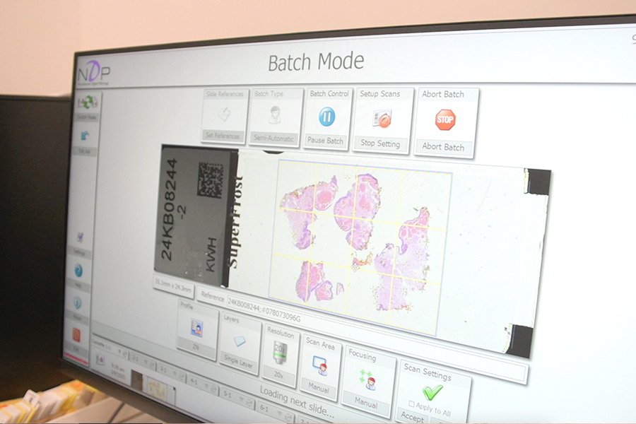
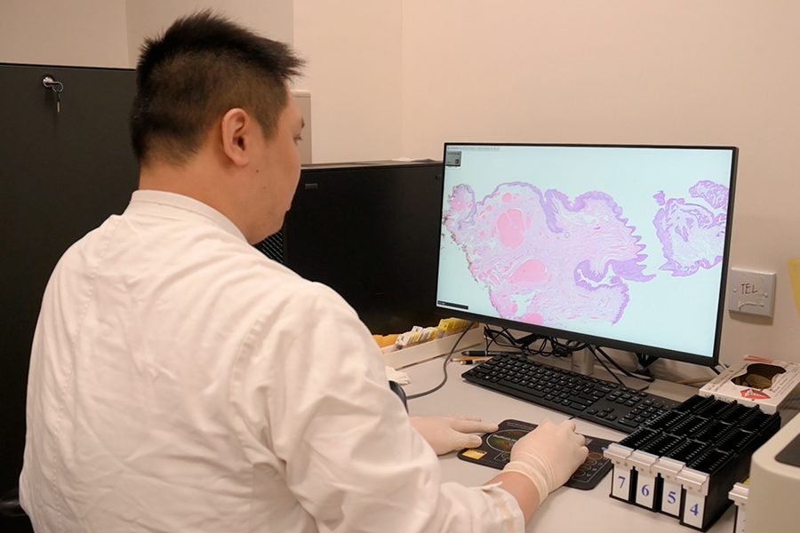
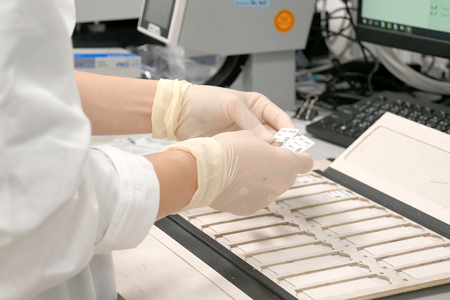
Tissue biopsy analysis is the key to cancer diagnosis and must be performed by experienced anatomical pathologists. However, manpower constraint is often a bottleneck. With the generous donation from the Tung Wah Group of Hospitals, the centre will purchase four scanners to convert traditional glass slide biopsy into digital image. The AI programme will analyse the images around the clock, locking onto suspected pathological changes to assist pathologists in diagnosis.
Screening for cervical and prostate cancer cases
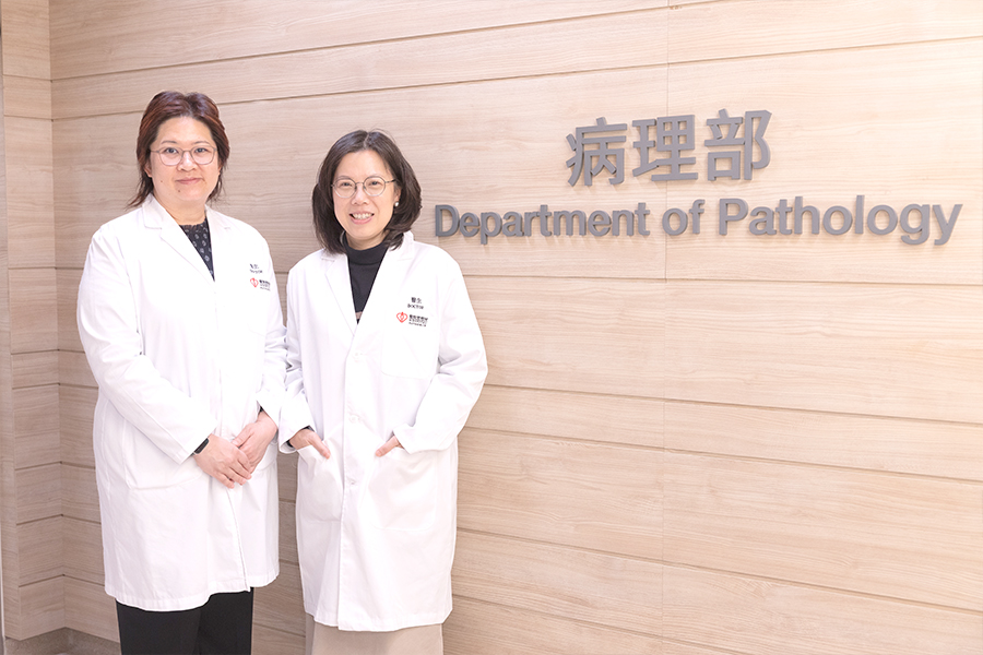
The new technology is expected to be applied to cervical smear and prostate cancer diagnoses by 2027. Dr Alice Chan, Consultant (Anatomical Pathology) of Department of Pathology at KWH, notes that in 2024 alone, the hospital processed over 26,000 cervical smears, with about 150 identified as pre-cancerous lesions. There were also over 5,024 prostate core biopsies, with at least 1,700 confirmed as cancer. She explains that pathologists previously had to manually review the samples one by one, AI now identifies high-risk cases for pathologists’ priority review. It achieves up to 90% accuracy, and shortens the testing time. “Samples that once took days to be reviewed can now be assessed on the very first day after they are flagged by the AI programme,” says Dr Chan.“In the past, cancer treatment options were limited. But with the development of targeted therapy and immunotherapy, diagnosis requires greater precision on the analysis of cells and biomarkers. Alongside an increased in public’s awareness, many tiny tumours are being detected earlier,” explains Dr Cindy Tse, Chief of Service of Department of Pathology at KWH. “AI enhances the efficiency of screening and diagnosis, allowing patients to receive appropriate treatment, and improving survival rates.” Dr Tse hopes the AI programme can benefit more patients.
Remote analysis of intraoperative frozen sections
The New Territories East Cluster also introduced remote frozen service by applying digital pathology. Previously, pathologists from North District Hospital had to travel to the Alice Ho Miu Ling Nethersole Hospital to examine the frozen section during a parathyroidectomy operation. Now, with the support of trained laboratory staff and real-time video, pathologists can analyse whole slide digital image of the frozen sections, to make immediate diagnoses remotely and save travel time. This technology will be extended to ‘margin biopsy’ for skin cancer patients.