Robot-assisted minimally invasive brain surgery brings hope to epilepsy patients
 Epilepsy is one of the most common brain disorders in Hong Kong. During seizure attack, the patient may suddenly collapse, lose consciousness with jerking movements of the arms and legs and foaming at the mouth. Epilepsy is like a time bomb, bringing a serious impact on patients’ daily life. Some patients’ epilepsy may be fairly controlled with medication, however, about 30% of the patients may have resistant epilepsy and may need to consider for surgery. Traditional invasive electroencephalogram (EEG) monitoring requires open-brain surgery and the treatment procedure is complicated and time-consuming. Some patients are often scared and hesitant to consider surgery.
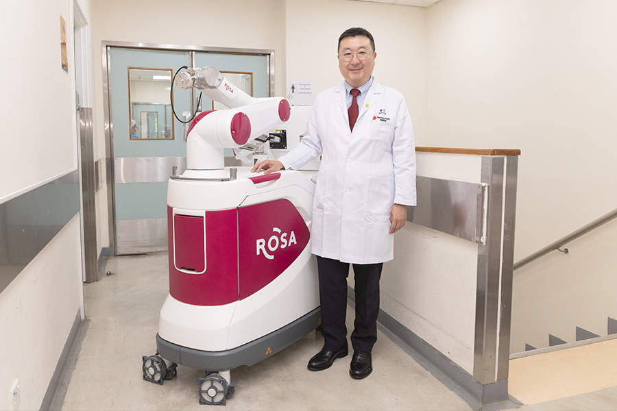
Queen Mary Hospital (QMH) has introduced fully automatic Robotic Stereotactic Assistance (Robot) since 2024 to assist minimally invasive brain surgery, opening a new direction in epilepsy treatment and allowing patients to glimpse the light of recovery. The Robot uses 3D imaging planning software to map the patient’s brain structure and then the surgeon can plan the surgical targets and trajectory. The robotic arm then automatically navigates and accurately locates the planned targets during the surgery. Compared with the traditional techniques such as the frame-based stereotaxy, surgeons now do not need to manually adjust the angle etc., reducing human errors during operation. At the same time, it can save about half of the operation time, lowering the surgical risk for the patients and improving overall efficiency.
Patients rediscover the beauty of life
“Stereoelectroencephalography surgery is to identify the focus of seizures. In the operating theatre, with the assistance of the Robot, multiple electrodes are inserted into different areas of the patient’s brain. The patient will then need to stay in the hospital for observation for several weeks to record the EEG,” says Dr Benedict Taw, Consultant of the Department of Neurosurgery. “When epilepsy occurs, the brain will emit abnormal brain signals. Doctors may be able to find the source of epilepsy according to the electrical recordings. Next, doctors will use radiofrequency thermocoagulation with the electrodes to emit high energy to destroy brain cells at the lesions. The treatment can help reduce epileptic seizures and lessen their impact on patients’ daily lives. Around 10% of patients may be cured of their seizures after thermocoagulation. Thermocoagulation also allows doctors to diagnose and confirm where the epileptic focus is, thereby assisting them with following up on the patient’s condition. If the seizures recur, further curative surgery may be offered.”“Epilepsy has a profound impact on patients’ daily lives. With the introduction of the new technology, patients can now undergo minimally invasive surgery to treat and manage their condition, sparing them the psychological burden of traditional open-brain surgery. Some patients have successfully regained their independence after surgery. They step onto the path of recovery and hope,” says Dr Ian Leung, Resident Specialist of the Department of Medicine.
 The team has introduced new technology for pre-operative examinations for epilepsy. Purely with MRI imaging, it is rather difficult for radiologists to accurately detect the area of the lesion. Now, with the introduction of voxel-based morphometry, the patient's MRI images are compared with the data in the database to deduce the range of abnormalities, allowing radiologists to estimate the range of lesions more effectively and accurately before surgery.
The Robot has already assisted in more than ten multidisciplinary surgeries involving epilepsy or movement disorders. The team plans to apply the Robot to the treatment of brain tumors and other brain conditions. Through the ongoing advancement of medical innovation, we sow the seeds of hope for patients.