Intraoperative MRI spares brain tumor patients a second craniotomy
 Brain surgery is considered one of the most challenging surgical procedures. In the past, when neurosurgeons performed traditional brain tumor resections, they primarily relied on pre-operative medical images and their past experience to determine the scope of resection. Patients had to undergo another magnetic resonance imaging (MRI) scan one or two months after surgery to confirm if all tumor cells had been completely removed. If residual tumor was found, the patient would need to undergo a second craniotomy, exerting a significant physical and psychological burden on the patient.
A real-time ‘map’ of the brain to locate tumor precisely
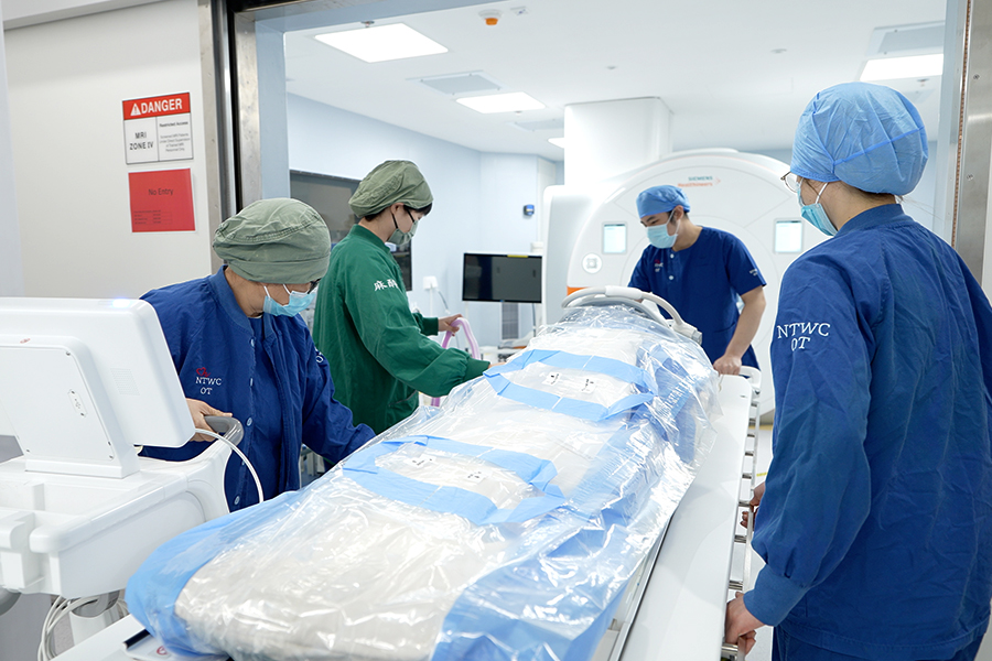
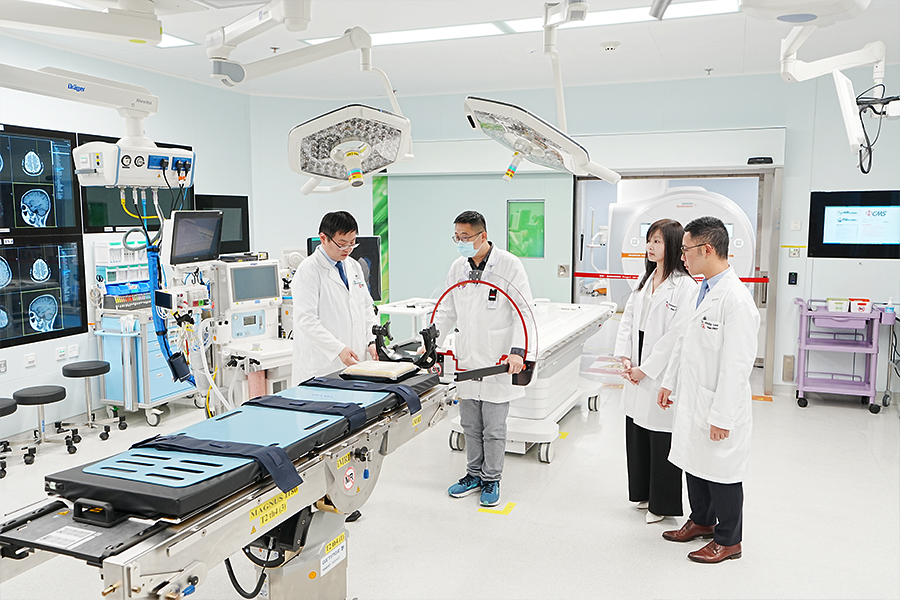
In March of this year, Tuen Mun Hospital (TMH) Neuroscience Centre launched Hong Kong's first Intraoperative Magnetic Resonance Imaging (IoMRI) Service. Now, after neurosurgeons resecting tumors, the team immediately transfers the patient to the adjacent MRI suite for an MRI scan. A radiologist immediately analyses the MRI images to precisely locate any small residual tumor. After returning to the operating theatre, neurosurgeons can assess whether a more thorough removal of tumor is necessary, preventing the spread of residual tumors while preserving as much surrounding healthy brain tissue as possible. The entire process is completed under a single session of anaesthesia, sparing patients the risk of a second surgery and greatly enhancing medical outcomes.TMH Consultant Radiologist Dr Philip Lee Yat-sing states: “Since the service was launched, the team has successfully performed surgery on 11 patients. All cases have achieved gross total resection of tumors based on preliminary results, realising our goal of removing brain tumors in a single operation.”
Double metal screenings to ensure MRI safety
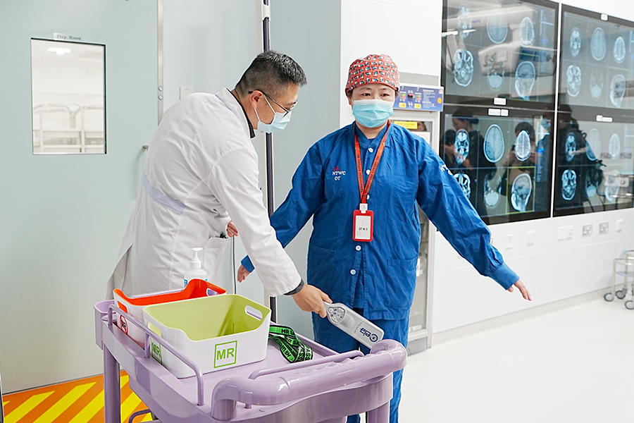
To ensure the entire process is flawless, the team conducted thorough preparations and drills before the service was launched. Referencing the concept of Crew Resource Management in aviation industry, they implemented a training programme and actively coordinated with multiple departments, including Neurosurgery, Anaesthesia and Operating Theatre Services, Radiology, and Nursing, to jointly establish a set of rigorous standard procedures and safety checklists.A few months ago, it was reported that in the United States, a man wearing a heavy metallic necklace entered an MRI suite and was sucked into the operating MRI machine, resulting in fatal injuries. TMH Deputy Chief of Service of the Department of Anaesthesia and Operating Theatre Services Dr Carmen Lam says: “We began strengthening our colleagues’ awareness of MRI safety since 2022. All medical staff entering the operating theatre must complete an MRI safety training course and obtain certification before receiving a red pass, which signifies they can enter the surgical area. Before entering the MRI suite, colleagues must undergo double checks on metal safety to ensure there are no metal items on or in their bodies, eliminating any potential hazards and protecting everyone’s safety.” The MRI safety standards established by TMH Neuroscience Centre provide an important reference for other hospitals implementing similar services in the future.
 TMH plans to gradually expand this new service to other complex brain surgeries within the year, such as paediatric brain tumors and invasive gliomas, and expects to serve 80 to 100 cases annually, allowing more patients to benefit.