MR technology improves the precision of surgeries
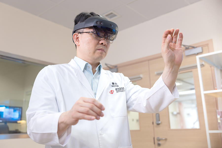 Head-mounted devices have become a popular electronic product in recent years. They can be used to watch films, play games, and even for medical applications. Since 2023, Prince of Wales Hospital (PWH) has applied mixed reality (MR) technology in orthopaedic tumor surgery, blending virtual medical images with the real clinical environment. When surgeons wear the device, they are able to see through the patient’s skin. The tissues underneath become clearly visible.
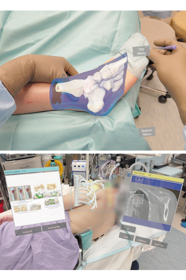
With traditional methods, surgeons could only make skin incisions in operation based on 2D images and clinical experience. Virtual images are overlaid onto the patient using MR technology, showing clearly the location of tumors, muscles, blood vessels, bones, and nerves. Surgeons can intuitively see the actual size and the depth of each tissue layer, understanding the relevance between different tissues. It directly guides pre-operative assessment, making the operation more accurate.
Accurate skin incisions to avoid amputation
This technology is mainly used in orthopaedic tumor surgeries at present. “Bone and soft tissue tumor surgeries are complex. Surgeons can carefully examine the exact locations of tumor and nearby vital structures, e.g., vessels and nerves with MR glasses. This allows them to predict the areas that need to be handled carefully. As a result, surgeons can understand the surgery more easily and locate the incision more quickly during operation, improving its precision and reducing the risk,” says Dr Wong Kwok-chuen, Consultant of PWH’s Department of Orthopaedics and Traumatology.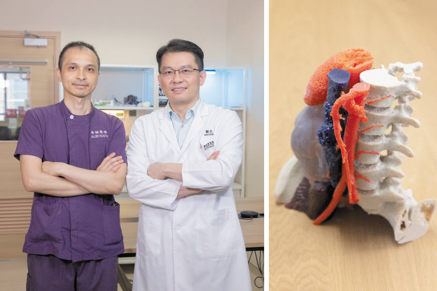 “Moreover, in treatments for primary bone cancers, surgeons can master every surgical step with the help of MR technology and other existing tools, e.g., computer navigation or 3D printing. It ensures the complete removal of bone cancers while increasing the chance of saving patients’ limbs and even their joints, avoiding limb amputation and retaining limb function.”
The preparation of MR technology is relatively simple. The MR glasses can be used within a few hours after uploading medical images to the system. The device can be used in an operating theatre after a basic disinfection. Surgeons can put on the glasses and 3D images will be projected with a simple gesture. Different interfaces can be switched with just a movement, allowing instant interaction. Not only can digital medical data be accessed instantly to support surgery, but the images in front of the surgeon’s eyes can also be captured for recording or sharing without interrupting the surgical procedure.
Surgeons in the operating theatre can consult external doctors, even overseas, by sharing surgical images and video calls, enabling immediate feedback. This new technology also facilitates remote learning, helping students to gain a clearer and more direct understanding of surgical procedures. Additionally, the recorded videos and images can also be used for clinical research.
“We have spent four years to apply the new technology to clinical practice. During the preparation, we conducted many tests and worked closely with the Information Technology Department to open access to the security system. We believe that more new technologies will be applied to different clinical services in the future, bringing new directions to patients' treatment plans,” says Dr Wong. The team is actively introducing new MR glasses for use during surgery. Other specialties including cardiothoracic surgery and ear, nose and throat are also accessing the feasibility of applying MR technology in pre-operative assessment.
Demonstration video of MR technology